

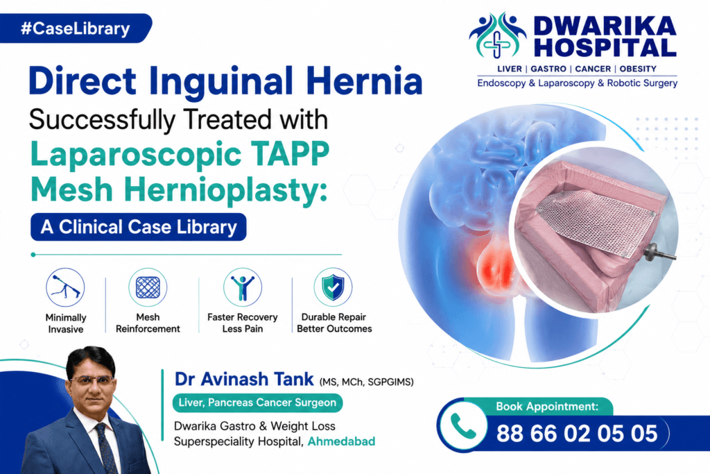
Knowledge Hub Layer: Clinical Case Library (Layer 4)
Suggested URL
/case-library/laparoscopic-tapp-repair-for-large-right-direct-inguinal-hernia/
Overview
A 69-year-old gentleman presented with a gradually enlarging swelling in the right groin associated with intermittent discomfort and heaviness for approximately three months. The swelling became more prominent during prolonged standing, walking, coughing, and straining and reduced spontaneously while lying down. There were no symptoms suggestive of bowel obstruction such as vomiting, abdominal distension, or inability to pass stool.
Clinical examination strongly suggested a right inguinal hernia. Ultrasonography confirmed a right inguinal hernia containing bowel loops with an approximately 29 mm fascial defect. A small contralateral (left) inguinal hernia was also demonstrated only during straining. The patient had significant cardiac history, including previous coronary angioplasty, making careful preoperative assessment essential. Cardiology evaluation confirmed preserved left ventricular systolic function (ejection fraction approximately 55%), and the patient was considered fit for elective surgery after optimization.
Considering the patient’s symptoms, progressive enlargement of the hernia, and the lifelong risk of incarceration and strangulation, elective laparoscopic Transabdominal Preperitoneal (TAPP) mesh hernioplasty was recommended.
During surgery, a large right direct inguinal hernia measuring approximately 5 cm with omentum as its contents was identified. The hernia was successfully reduced, a large polypropylene mesh was placed within the preperitoneal space, and the peritoneum was closed without intraoperative complications. The patient’s postoperative recovery was uneventful, with early mobilization, pain control, and discharge after recovery.
This case demonstrates the importance of timely diagnosis, appropriate patient selection, meticulous laparoscopic surgical technique, and comprehensive perioperative assessment in elderly patients with inguinal hernia.
Case Highlights
| Parameter | Details |
|---|---|
| Age | 69 years |
| Gender | Male |
| Primary Complaint | Right groin swelling with discomfort |
| Duration | Approximately 3 months |
| Hernia Side | Right |
| Hernia Type | Direct inguinal hernia |
| Hernia Size | Approximately 5 cm intraoperatively |
| Hernia Contents | Greater omentum |
| Contralateral Hernia | Small left inguinal hernia seen only on straining |
| Imaging | Ultrasonography |
| Surgical Procedure | Laparoscopic Right TAPP Mesh Hernioplasty |
| Mesh Used | Polypropylene mesh (10 × 15 cm) |
| Fixation | Absorbable tack fixation |
| Anaesthesia | General anaesthesia |
| Intraoperative Complications | None |
| Postoperative Course | Uneventful |
| Outcome | Successful repair with good early recovery |
Values are based on the uploaded operative records, ultrasound report, and perioperative documentation.
Patient Presentation
How did the patient first present?
The patient presented with a progressively increasing swelling in the right groin over approximately three months. Initially, the swelling was small and appeared only after physical activity. Gradually, it became more noticeable and produced a sensation of heaviness while standing or walking.
He reported:
- Right groin swelling
- Mild discomfort rather than severe pain
- Increase in swelling while standing
- Enlargement during coughing and straining
- Reduction on lying down
- No fever
- No vomiting
- No bowel obstruction
- No gastrointestinal bleeding
- No urinary obstruction
The symptoms were beginning to interfere with routine daily activities and quality of life, prompting surgical consultation.
Relevant Medical History
The patient’s medical history was significant for:
- Coronary artery disease
- Previous coronary angioplasty
- Hypertension
- Diabetes mellitus (HbA1c approximately 6.8%)
- Previous hospitalization for coronavirus infection (documented history)
- Long-term treatment with cardiovascular medications
Despite these comorbidities, the patient remained functionally active and independent in activities of daily living.
Physical Examination
Clinical examination demonstrated findings consistent with a right inguinal hernia:
- Visible right groin swelling
- Swelling increased with cough impulse
- Reducible hernia
- No signs of skin inflammation
- No features of strangulation
- Abdomen soft and non-distended
The absence of irreducibility, severe tenderness, fever, tachycardia, or intestinal obstruction suggested that this was an uncomplicated but symptomatic inguinal hernia suitable for elective repair.
Clinical Evaluation
How was the diagnosis suspected?
The diagnosis was primarily based on a combination of the patient’s characteristic symptoms and focused physical examination.
Several clinical features strongly favored an inguinal hernia:
- Groin swelling that increased during standing
- Enlargement with coughing or straining
- Reduction while lying down
- Positive cough impulse
- Absence of inflammatory signs
These findings made an inguinal hernia the leading diagnosis.
Differential Diagnosis Considered
Although the clinical findings strongly suggested an inguinal hernia, other causes of groin swelling were considered during evaluation:
- Indirect inguinal hernia
- Femoral hernia
- Lipoma of the spermatic cord
- Enlarged inguinal lymph nodes
- Saphena varix
- Hydrocele of the cord
- Soft tissue tumors
- Femoral artery aneurysm (rare)
Ultrasonography was performed to confirm the diagnosis, define the anatomy, identify the hernia contents, and detect any occult contralateral hernia before surgery.
5. Investigations and Imaging Findings
Why were investigations necessary?
Although inguinal hernia is primarily a clinical diagnosis, investigations were performed in this patient for three important reasons:
- To confirm the diagnosis and define the anatomy.
- To assess the patient’s fitness for surgery, considering his age and multiple medical comorbidities.
- To identify factors that could influence operative planning and perioperative risk.
Baseline Blood Investigations
Routine preoperative investigations demonstrated satisfactory organ function for elective laparoscopic surgery.
Hematology
| Investigation | Result | Clinical Interpretation |
|---|---|---|
| Hemoglobin | 12.0 g/dL | Mild anemia but acceptable for elective surgery |
| Total WBC Count | 6,560/µL | No evidence of active infection |
| Platelets | 213,000/µL | Normal coagulation reserve |
Peripheral smear demonstrated normocytic normochromic red blood cells with eosinophilia but no evidence of acute hematological abnormality.
Renal Function
Renal parameters remained within acceptable limits.
- Serum Creatinine: 0.88 mg/dL
- Sodium: 138.3 mmol/L
- Potassium: 4.2 mmol/L
- Chloride: 105.6 mmol/L
Normal renal function allowed safe administration of general anaesthesia and perioperative medications.
Liver Function
The liver profile was normal.
- Bilirubin: Normal
- SGOT: Normal
- SGPT: Normal
- Alkaline phosphatase: Normal
- Albumin: Normal
These findings indicated preserved hepatic function and adequate protein reserve for postoperative wound healing.
Diabetes Assessment
The patient had known diabetes mellitus.
HbA1c measured approximately 6.8%, indicating reasonably controlled diabetes before surgery.
Adequate glycaemic control is important because poorly controlled diabetes increases the risk of:
- Surgical site infection
- Delayed wound healing
- Mesh infection
- Longer hospital stay
- Hernia recurrence
Appropriate perioperative glucose control was therefore incorporated into the surgical plan.
Coagulation Profile
The coagulation profile was satisfactory.
- PT
- INR
- APTT
No coagulation abnormality requiring correction before surgery was documented.
Ultrasonography of the Abdomen and Groin
Why was ultrasound performed?
Ultrasonography was requested to:
- Confirm the diagnosis
- Determine the side of the hernia
- Identify hernia contents
- Measure the fascial defect
- Detect occult contralateral hernia
- Exclude other causes of groin swelling
Ultrasound Findings
The examination demonstrated:
- Right inguinal hernia
- Herniation of bowel loops
- Fascial defect measuring approximately 29 mm
- Left inguinal hernia seen only during straining
- No intra-abdominal mass
- No ascites
- Normal liver
- Normal gallbladder
- Normal pancreas
- Normal spleen
- Normal kidneys
- Normal urinary bladder
These findings confirmed bilateral inguinal weakness, although only the right side was clinically symptomatic.
Cardiac Evaluation
Because of the patient’s previous coronary angioplasty and cardiovascular disease, detailed preoperative cardiac evaluation was performed.
ECG
Electrocardiography showed sinus rhythm without acute ischemic changes that would preclude elective surgery.
Echocardiography
Important findings included:
- Left ventricular ejection fraction approximately 55%
- Normal left ventricular systolic function
- No regional wall motion abnormality
- Mild diastolic dysfunction
- Mild aortic valve sclerosis
- No pulmonary hypertension
Overall, the cardiologist considered the patient suitable for elective surgery after optimization.
How Did These Investigations Influence Management?
Each investigation contributed to surgical planning.
| Investigation | Impact on Decision-Making |
|---|---|
| Blood tests | Confirmed fitness for surgery |
| HbA1c | Guided perioperative diabetic management |
| Ultrasound | Confirmed right direct inguinal hernia and identified contralateral weakness |
| ECG | Excluded unstable cardiac disease |
| Echocardiography | Confirmed acceptable cardiac function for general anaesthesia |
Together, these investigations allowed safe planning of elective laparoscopic repair rather than emergency intervention.
6. Final Diagnosis
Final Clinical Diagnosis
Large symptomatic right direct inguinal hernia with omental contents in a 69-year-old male, associated with an asymptomatic left inguinal hernia demonstrable only on straining.
Clinical Classification
The uploaded operative note confirms a direct inguinal hernia.
From an educational perspective, direct inguinal hernias:
- protrude through Hesselbach’s triangle,
- occur medial to the inferior epigastric vessels,
- result from weakness of the posterior wall of the inguinal canal,
- are more common in elderly men because of attenuation of the transversalis fascia.
Unlike indirect inguinal hernias, direct hernias usually do not traverse the deep inguinal ring.
Why Was This Considered a Large Hernia?
Although preoperative ultrasonography measured the fascial defect at approximately 29 mm, the operative findings demonstrated a large direct hernia measuring approximately 5 cm, indicating that intraoperative assessment provided a more accurate estimate of the defect size.
7. Why Surgery Was Recommended
Why not simply observe?
Observation (“watchful waiting”) may be appropriate for carefully selected patients with minimally symptomatic inguinal hernias.
However, surgery was recommended in this patient because several factors favored definitive repair.
Progressive Symptoms
The patient reported:
- Increasing groin swelling
- Progressive heaviness
- Discomfort during daily activities
These symptoms suggested gradual enlargement of the hernia.
Risk of Complications
Untreated inguinal hernias may progressively enlarge and eventually develop complications such as:
- Incarceration
- Intestinal obstruction
- Strangulation
- Bowel ischemia
- Need for emergency surgery
Elective repair carries significantly lower morbidity than emergency hernia surgery.
Better Functional Recovery
Elective laparoscopic repair generally offers:
- Less postoperative pain
- Earlier walking
- Faster return to normal activity
- Lower wound infection rate
- Excellent long-term outcomes when performed using modern mesh techniques
Patient Factors
Despite his age and previous coronary angioplasty, the patient demonstrated:
- Good functional status
- Controlled diabetes
- Acceptable cardiac function
- Adequate physiological reserve
Following multidisciplinary assessment, elective surgery was considered safer than delaying treatment.
8. Surgical Planning
Why was a laparoscopic approach selected?
After reviewing the clinical findings, imaging, and overall health status, laparoscopic Transabdominal Preperitoneal (TAPP) repair was selected.
Advantages of TAPP Repair
The laparoscopic approach offers several advantages:
- Excellent visualization of both groins
- Ability to identify occult contralateral hernias
- Wide preperitoneal mesh placement
- Less postoperative pain
- Smaller incisions
- Earlier mobilisation
- Faster return to routine activities
- Lower chronic groin pain compared with some open repairs
In this patient, laparoscopy also allowed confirmation of the anatomy and facilitated repair of a large direct defect under magnified vision.
Mesh Planning
Based on the anticipated size of the myopectineal orifice and the large direct defect, a 10 × 15 cm polypropylene mesh was selected.
Large mesh overlap beyond the defect is important to reduce recurrence by reinforcing the entire myopectineal orifice rather than simply closing the hernia opening.
9. Surgical Challenges
Although this was an uncomplicated elective case, several factors required careful operative planning.
Large Direct Hernia
Large direct hernias often have:
- Broad defects
- Thin transversalis fascia
- Significant bulging
- Attenuated posterior wall
These characteristics increase the importance of obtaining wide mesh coverage.
Omental Herniation
The operative note documented omentum as the hernia content.
Reduction of bulky omentum requires gentle traction to avoid bleeding or injury.
Elderly Patient with Cardiac Disease
Previous coronary angioplasty required:
- Careful anaesthetic planning
- Cardiovascular monitoring
- Appropriate perioperative medication management
- Early postoperative mobilisation to reduce cardiopulmonary complications
Prevention of Recurrence
Successful repair depends not only on reducing the hernia but also on:
- Adequate preperitoneal dissection
- Complete exposure of Cooper’s ligament
- Wide mesh overlap
- Secure fixation where indicated
- Proper closure of the peritoneal flap
These principles reduce recurrence and chronic postoperative pain.
10. Operative Strategy
Which operation was performed?
The patient underwent Laparoscopic Right Transabdominal Preperitoneal (TAPP) Mesh Hernioplasty under general anaesthesia. According to the operative note, a 10 × 15 cm polypropylene mesh was placed in the preperitoneal space and secured with absorbable tacks. The peritoneal flap was then closed over the mesh. No intraoperative complications were documented.
Why was the TAPP approach chosen?
The TAPP technique allows the surgeon to enter the peritoneal cavity, create a preperitoneal plane, reduce the hernia, and reinforce the entire myopectineal orifice with a large mesh. It offers several advantages:
- Excellent visualization of groin anatomy
- Easy identification of direct, indirect, and femoral defects
- Detection of occult contralateral hernias
- Wide mesh coverage of all potential hernia sites
- Reduced postoperative pain
- Faster recovery and earlier return to normal activities
For this patient, the laparoscopic approach provided optimal visualization of the large direct defect while minimizing surgical trauma.
Step-by-Step Operative Procedure
1. Creation of Pneumoperitoneum
Pneumoperitoneum was established using the open technique, and laparoscopic ports were inserted under direct vision.
2. Diagnostic Laparoscopy
The abdominal cavity was inspected to confirm the diagnosis and identify the hernia anatomy.
A large right direct inguinal hernia was visualized.
3. Reduction of Hernia Contents
The hernia contained greater omentum, which was gently reduced back into the abdominal cavity.
Careful atraumatic handling minimized bleeding and prevented omental injury.
4. Development of the Preperitoneal Space
A peritoneal incision was made above the hernia defect.
The peritoneal flap was carefully developed to expose:
- Pubic symphysis
- Cooper’s ligament
- Inferior epigastric vessels
- Myopectineal orifice
Adequate exposure of these landmarks is fundamental for a durable hernia repair.
5. Reduction of the Direct Hernia Sac
The attenuated transversalis fascia forming the direct hernia sac was identified.
The sac was carefully dissected and reduced from the hernia defect.
6. Mesh Placement
A 10 × 15 cm polypropylene mesh was introduced into the preperitoneal space.
The mesh was positioned to provide generous overlap of:
- Direct space
- Indirect space
- Femoral canal
- Obturator region
This broad reinforcement decreases the risk of future recurrence.
7. Mesh Fixation
The operative note documents fixation of the mesh using absorbable tacks.
Appropriate fixation helps prevent mesh migration during early healing while minimizing the long-term risk of chronic groin pain.
8. Closure of the Peritoneal Flap
After confirming satisfactory mesh positioning, the peritoneal flap was closed with absorbable tacks, completely separating the mesh from the abdominal viscera.
This reduces the risk of bowel adhesion to the prosthetic mesh.
9. Completion of Surgery
The operative field was inspected for:
- Hemostasis
- Mesh position
- Integrity of the peritoneal closure
No intraoperative complications were reported.
The ports were removed, and the skin was closed with staples.
11. Intraoperative Findings
What was found during surgery?
The operative findings were consistent with the preoperative diagnosis but provided additional anatomical details.
Major Findings
- Large right direct inguinal hernia
- Defect measuring approximately 5 cm
- Hernia contents consisted of greater omentum
- No bowel ischemia
- No strangulation
- No incarcerated bowel
- Successful reduction of hernia contents
- No unexpected intra-abdominal pathology
These findings confirmed that elective laparoscopic repair was appropriate and technically feasible.
Clinical Importance of These Findings
The discrepancy between the ultrasound defect (29 mm) and the operative estimate (~5 cm) illustrates an important surgical principle: laparoscopy provides direct visualization of the entire myopectineal orifice and often reveals the true extent of a direct hernia more accurately than preoperative imaging.
The presence of omentum rather than bowel also reduced the immediate risk of bowel injury during reduction.
12. Mesh Placement and Surgical Reconstruction
Unlike many abdominal operations, inguinal hernia repair does not involve removal of tissue or reconstruction of the gastrointestinal tract.
Instead, the operation restores the strength of the abdominal wall by reinforcing the weakened posterior wall of the inguinal canal.
Why was mesh used?
Modern international guidelines recommend mesh repair for most adult inguinal hernias because it:
- Significantly reduces recurrence
- Provides durable reinforcement
- Allows tension-free repair
- Improves long-term outcomes
The 10 × 15 cm polypropylene mesh used in this case provided broad coverage of the entire myopectineal orifice, rather than simply covering the visible defect.
13. Postoperative Care
Immediate Recovery
Following surgery, the patient was transferred to the recovery area for routine monitoring.
Postoperative management included:
- Observation of vital signs
- Pain assessment
- Monitoring for urinary retention
- Early oral intake after recovery from anaesthesia
- Early mobilization
- Deep vein thrombosis prevention
The postoperative orders documented analgesia, antibiotics, intravenous fluids, and monitoring of urine output and vital signs.
Pain Management
Effective pain control facilitates:
- Early walking
- Improved breathing
- Reduced pulmonary complications
- Earlier discharge
The minimally invasive approach generally results in less postoperative pain than conventional open surgery.
Early Mobilization
The patient was encouraged to:
- Sit up soon after surgery
- Walk on the day of surgery or the following morning
- Resume light daily activities as tolerated
Early mobilization reduces the risk of:
- Deep vein thrombosis
- Pulmonary complications
- Muscle deconditioning
Wound Care
The laparoscopic port sites were closed with staples.
Patients are advised to:
- Keep wounds clean and dry
- Watch for redness or discharge
- Attend scheduled follow-up for wound assessment and staple removal, if required
14. Outcome and Recovery
Early Outcome
The operation was completed successfully without intraoperative complications.
The patient experienced:
- Relief of the groin swelling
- Stable postoperative recovery
- No documented surgical complications
- Good tolerance of oral intake
- Early mobilization
Expected Functional Recovery
Most patients undergoing uncomplicated laparoscopic TAPP repair can expect:
- Walking within hours of surgery
- Return to routine household activities within a few days
- Return to office work within 1–2 weeks
- Gradual resumption of strenuous exercise after 4–6 weeks, depending on clinical assessment
15. Long-Term Follow-up
Standard postoperative surveillance after laparoscopic inguinal hernia repair includes:
- Assessment of wound healing
- Evaluation for chronic groin pain
- Detection of seroma or hematoma
- Monitoring for recurrence
- Return to normal activities
- Advice regarding gradual lifting and exercise
Patients should seek medical review if they develop:
- Persistent groin swelling
- Increasing pain
- Fever
- Wound infection
- New groin bulge suggestive of recurrence
16. Timeline of Care
| Timeline | Clinical Event |
|---|---|
| Initial symptoms | Progressive right groin swelling and heaviness |
| Clinical evaluation | Reducible right inguinal hernia suspected |
| Ultrasound | Right inguinal hernia with bowel loops; left hernia on straining |
| Cardiac assessment | Fitness confirmed after previous angioplasty |
| MDT surgical planning | Elective laparoscopic repair recommended |
| Surgery | Laparoscopic Right TAPP Mesh Hernioplasty |
| Intraoperative findings | Large 5 cm direct hernia with omentum |
| Mesh repair | 10 × 15 cm polypropylene mesh placed |
| Recovery | Uneventful postoperative course |
17. Surgical Pearls
- Large direct inguinal hernias are best assessed under laparoscopic magnified vision.
- Wide preperitoneal dissection is essential for adequate mesh overlap.
- Cooper’s ligament should be clearly identified before mesh placement.
- Reduction of omental contents should be gentle to prevent bleeding.
- Reinforcement of the entire myopectineal orifice is more important than simply closing the defect.
- Proper peritoneal closure minimizes the risk of bowel adhesion to the mesh.
- Comprehensive preoperative optimization improves outcomes in elderly patients with cardiac comorbidities.
18. Learning Points
- Direct inguinal hernias are common in older men due to weakening of the posterior inguinal wall.
- Groin swelling that enlarges on coughing and reduces on lying down is highly suggestive of an inguinal hernia.
- Ultrasonography is useful when the diagnosis or anatomy is uncertain.
- Cardiac assessment is important before elective surgery in patients with coronary artery disease.
- Laparoscopic TAPP repair provides excellent visualization of groin anatomy.
- Large mesh overlap reduces recurrence rates.
- Elective repair is generally safer than emergency surgery for incarcerated hernias.
- Early mobilization reduces postoperative complications.
- Good diabetes control contributes to better wound healing.
- Long-term follow-up is important to detect recurrence or chronic pain.
19. Learning for Patients and Family
- A groin swelling should never be ignored, even if it disappears on lying down.
- Hernias do not heal with medicines, belts, or exercises alone.
- Elective surgery is usually safer than waiting for an emergency.
- Laparoscopic surgery often results in smaller scars, less pain, and quicker recovery.
- Follow your surgeon’s instructions regarding lifting heavy weights after surgery.
- Maintain a healthy body weight and avoid chronic constipation or persistent coughing, which increase abdominal pressure.
- Attend all scheduled follow-up visits.
- Seek immediate medical attention if the swelling becomes painful, irreducible, or is associated with vomiting.
20. Learning for Referring Doctors and Primary Care Physicians
- Progressive groin swelling with a positive cough impulse strongly suggests an inguinal hernia.
- Differentiate direct, indirect, and femoral hernias through careful clinical examination and imaging when needed.
- Early referral is advisable for symptomatic or enlarging hernias.
- Assess cardiovascular risk and optimize comorbidities before elective surgery.
- Laparoscopic TAPP repair is particularly advantageous when bilateral defects or occult contralateral hernias are suspected.
- Prompt referral before incarceration or strangulation improves patient outcomes.
21. Frequently Asked Questions (FAQ)
1. What is a direct inguinal hernia?
A direct inguinal hernia occurs when abdominal contents bulge through a weakened area of the posterior wall of the inguinal canal (Hesselbach’s triangle). It is most commonly seen in older adults due to age-related weakening of the abdominal wall.
2. Why was surgery recommended?
The hernia was symptomatic, progressively enlarging, and carried a lifelong risk of incarceration and strangulation. Elective laparoscopic repair provides definitive treatment and reduces the risk of emergency complications.
3. What is laparoscopic TAPP hernia repair?
TAPP (Transabdominal Preperitoneal) repair is a minimally invasive procedure in which a mesh is placed behind the abdominal wall through small incisions to reinforce the weakened area.
4. Is mesh safe?
Modern polypropylene mesh has been extensively studied and is considered the standard of care for most adult inguinal hernias because it significantly reduces recurrence when used appropriately.
5. How long does recovery take?
Most patients resume light activities within a few days and return to routine work within 1–2 weeks. Heavy lifting should be avoided for several weeks, depending on the surgeon’s advice.
6. Can the hernia recur?
Although recurrence is uncommon after a properly performed mesh repair, it remains possible. Maintaining a healthy weight, avoiding smoking, treating chronic cough or constipation, and following postoperative advice help reduce the risk.
7. Can both sides be repaired laparoscopically?
Yes. One advantage of the TAPP approach is that both groins can be inspected, and bilateral hernias can often be repaired during the same operation when indicated.
8. When should I seek urgent medical attention?
Seek immediate medical care if the groin swelling becomes painful, cannot be pushed back, is associated with vomiting, abdominal distension, fever, or redness over the swelling, as these may indicate incarceration or strangulation.
22. Related Pages
Disease Knowledge Hub
- Direct Inguinal Hernia
- Inguinal Hernia
- Bilateral Inguinal Hernia
- Recurrent Inguinal Hernia
Treatment / Service Page
- Laparoscopic Inguinal Hernia Repair (TAPP)
- Laparoscopic Hernia Surgery
- Minimally Invasive Hernia Surgery
23. Educational Disclaimer
This clinical case is shared for educational purposes to illustrate the evaluation and management of a patient with a direct inguinal hernia. Patient-identifying information has been omitted to preserve confidentiality.
The investigations, treatment decisions, and surgical approach described in this report were based on the individual patient’s clinical condition and the treating surgeon’s judgment. Management may differ for other patients depending on age, symptoms, anatomy, associated illnesses, imaging findings, and overall health.
Patients should not use this case as a substitute for professional medical advice. Anyone with symptoms suggestive of an inguinal hernia should seek consultation with an experienced surgeon for a comprehensive evaluation and individualized treatment plan.
Was this article helpful?

Super-specialist GI, bariatric & cancer surgeon. SGPGIMS (India's premier GI centre) + advanced training in Japan & South Korea. Read full profile →
Last reviewed: July 2026


Comments
Be the first to share your thoughts on this article.